
Hematology is the study of the blood and diseases associated with blood. Blood is made up of 3 main cell types: red blood cells, white blood cells, and platelets. Red blood cells make up the majority of the blood cells and are responsible for housing a molecule known as hemoglobin which transports oxygen and carbon dioxide. White blood cells are immune cells within the blood that can travel throughout the body to sites of infection, injury, or disease. Platelets, along with many clotting factors, are responsible for forming blood clot. They are fragments of large cells produced by the bone marrow. Besides cells, the blood is composed of many minerals and electrolytes. All these cells, minerals, electrolytes, and other components are floating in water. For this reason, dehydration has a serious effect on blood volume. When a patient is severely dehydrated, water is pulled out of the blood to hydrate other cells making the blood more viscous and decreasing its ability to flow.
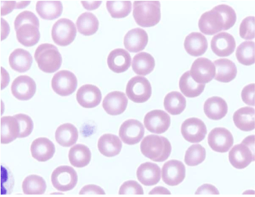
Red blood cells are very small cells that have a relatively short lifespan. In mammals, they do not have a nucleus, are biconcave, and spherical. These adaptations allow them to carry a large amount of hemoglobin (and in turn oxygen) at a time for efficient gas exchange. There are main different abnormal shapes that can be seen in red blood cells and these are a bit too complicated for this post, but each change is indicative of some disease process occurring in the body.
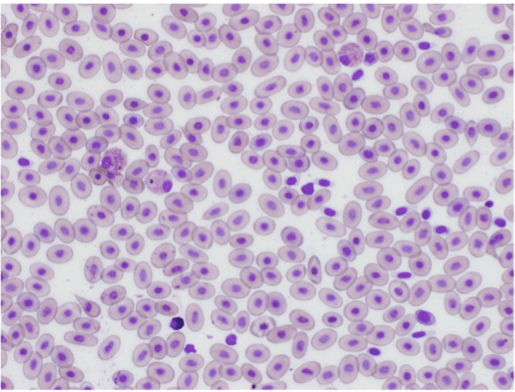
Fun fact #1: Animals in the Camelid family (llamas, alpacas, camels) have elliptical red blood cells.

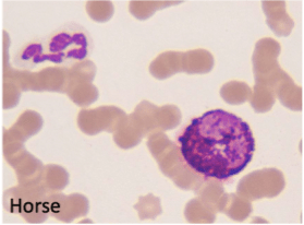
Fun fact #2: Birds, reptiles, and amphibians have very large elliptical shaped red blood cells. Their red blood cells also retain their nucleus. Continue reading to find out what issues this can cause when analyzing exotic animal blood!
Each white blood cell has a unique shape, size, and function within the immune system. They are responsible for attacking foreign material and disease. It is important for them to be located within the blood because the blood serves as a transport system throughout the entire body. They are often the first line of defense against foreign material because of the widespread distribution of the cells within the blood.
- Granulocytes: Granulocytes are named because they contain many granules within their cytoplasm. When these cells encounter something that they deem as a threat, they release their granules which will act on the invader to destroy it or mark it for further intervention.
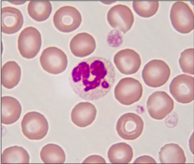
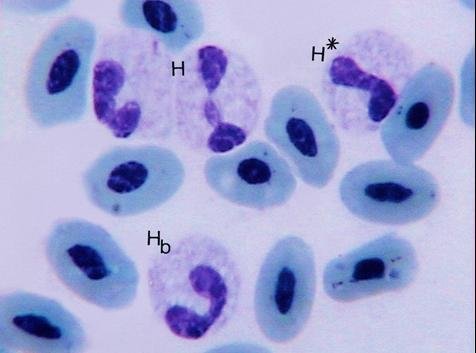
- Neutrophils: In mammals, neutrophils are the most abundant white blood cell in the blood. They are often the first cell to respond to inflammation and infection. They are also capable of “eating” foreign material like bacteria. Neutrophils are identified by their distinct segmented, purple nucleus. In some species, the body holds a large storage pool of neutrophils that it can dispense when needed. The presence of immature neutrophils in the blood indicates that the body’s immune system is overwhelmed by the insult and it cannot produce neutrophils fast enough to fight it. Immature neutrophils look slightly different from their mature counterparts. Instead of the distinct segmented nucleus, the nucleus has a more consistent thickness and sometimes resembles a horseshoe.

Fun fact #3: Birds have a cell called heterophils that resemble neutrophils and play the same role in immune function.
Fun fact #4: Neutrophils are not the most abundant white blood cell in reptiles. Continue reading to find out what is!
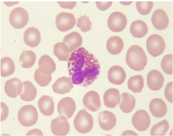
- Eosinophils: Eosinophils are the second most common granulocyte, but they are only present in very small numbers in normal blood. Eosinophils respond to allergic reactions, parasitic infections, and some weird diseases like fungal infections. Eosinophils are identified by their distinct bright pink granules.
Fun fact #5: Certain dog breeds, like greyhounds and golden retrievers, have grey eosinophil granules.
- Basophils: In most species, it is extremely rare to find any basophils in normal blood. Basophils play a similar role to eosinophils by responding to allergic reactions and parasites. Basophils resemble eosinophils but their granules are purple.
Fun fact #6: Rabbits, turtles, tortoises, and birds tend to have high numbers of basophils in health, sometimes making up 60% of the white blood cells!
- Mononuclear cells: Mononuclear cells are the white blood cells that have a round nucleus. These cells are often found later in infection or in chronic conditions.
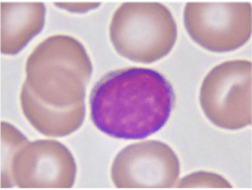
- Lymphocytes: These mononuclear cells are probably the most known type in humans as these are your B and T cells. They are usually the second most common type of white blood cell seen in healthy animals. They are seen post-vaccination, in chronic infectious diseases, autoimmune diseases, and endocrine diseases. They are similar in size to granulocytes, but the nucleus takes up almost the entire cell.
Fun fact #7: Lymphocytes are the most common white blood cell in adult cows, goats, sheep, rats, pigs, mice, and many bird species.
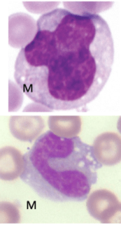
- Monocytes/Macrophages: Monocytes/macrophages are the largest of the white blood cells. They are called monocytes when they are still in the blood and macrophages when they enter the tissue. They are primarily responsible for “eating” foreign materials. When they “eat”, they present the foreign object to T cells so that they can determine how to respond. They are commonly seen in chronic, persistent infections when the body has depleted neutrophil stores. Their large size makes them easy to identify because they can have an extremely variable nucleus.
Blood indices are commonly used in both human medicine and veterinary medicine. There are two main ways to evaluate blood: manual and laboratory diagnostics. Both give a doctor valuable information about the composition and integrity of the blood and the cells. Manual diagnostics are readily available because they can be done by a clinician in practice. Most laboratory diagnostics will be sent off to a diagnostic lab that has specialized analyzers to determine values. Both methods are necessary to give a holistic view of what is going on in the blood and in the body.
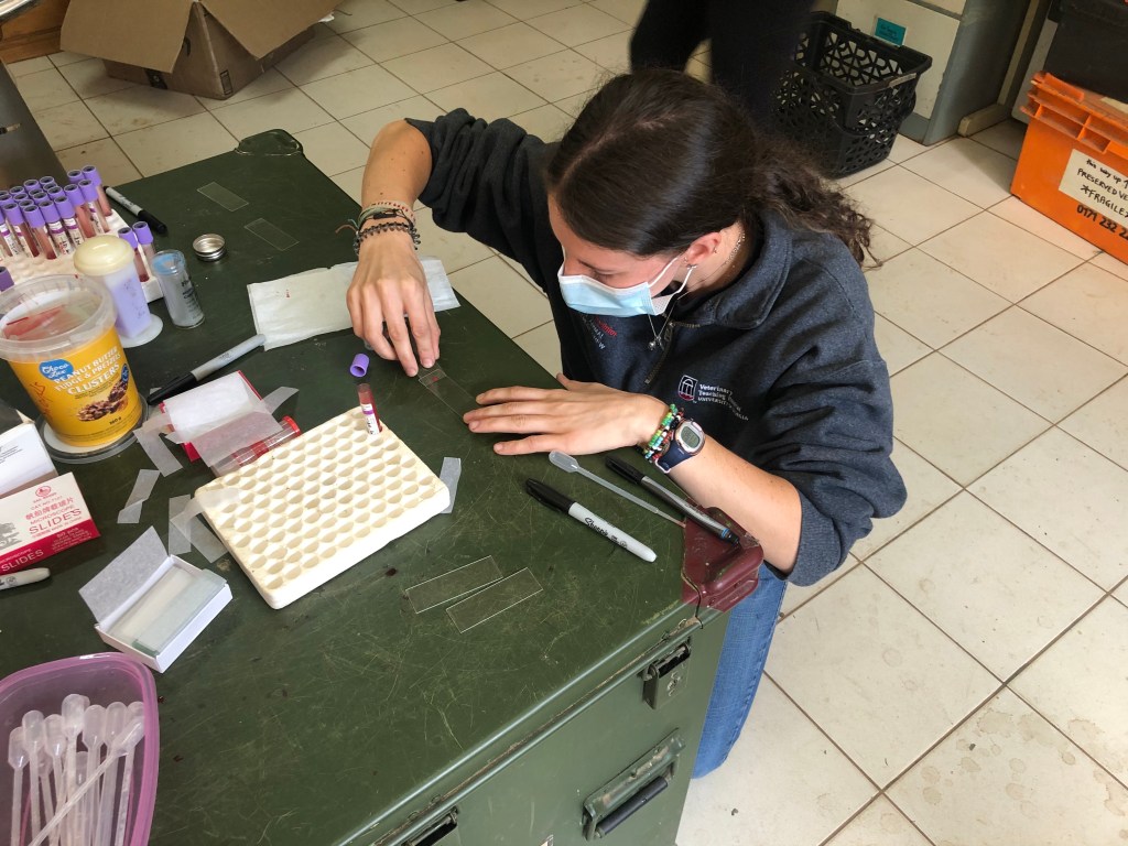
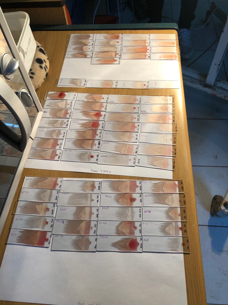
Manual analysis is typically done using a blood smear. Blood smears are very easy to do and require little supplies. All you need is a drop of unclotted blood, 2 slides, and stain. The goal is to spread out the blood cells into a thin layer on the slide so that you can evaluate number of each cell type and the shape, color, and size of the red blood cells. The most common stain used is called Diff-Quik which gives the distinct pink and purple coloration to the cells.
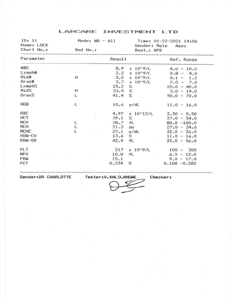
Laboratory analyses vary immensely based on what you are trying to test for and what lab you are using. The most common analyses performed are CBCs (complete blood counts) and Chemistry. Chemistry evaluates the electrolytes and minerals in the blood. I will not be going into detail on chemistry panels in this post, but it is important to know that this information can tell you a million things about what is going on in all parts of the body. CBCs will give you a count of the red blood cells, total white blood cells, number/percentage of each type of white blood cell, hemoglobin concentration, and some other information about your red blood cells.
Main issues associated with CBC results:
- High RBC count: A high red blood cell count is usually indicative of dehydration. As stated above, water is pulled out of the blood making it more concentrated with red blood cells. You should also see an increased hematocrit (HCT) and hemoglobin concentration because of the increased relative number of red blood cells. There could be an issue with destruction of your red blood cells, or the body is creating too many.
- Low RBC count: A low red blood cell count means that the patient is anemic. There are several different types of anemia and the other red blood cell indices (MCV, MCHC, RDW) can help you determine the cause of the anemia.
- High total WBC count: A high total white blood cell count usually indicates infection or inflammation. However, total WBC counts are usually not diagnostic on their own. The change in the level of each individual white blood cell can indicate a different type of response occurring in the body.
- Low total WBC count: A low total white blood cell count could indicate a bone marrow disease process preventing production of the cells. However, if one type of white blood cell is still high, bone marrow disease is unlikely because all cells would be affected. Other causes of immune suppression can cause decreased white blood cell production like steroid use, stress, or chemotherapy.
As you can see from the information above, species differences when it comes to blood analyses are very common. For this reason, you should never compare two sets of results from two different species and expect to see the same thing. To further complicate things when working with wildlife, there are very rarely normal reference ranges for these tests for a given species. We have normal ranges for our domestic species, so it is very easy to look at a lab report and determine what is abnormal. For our exotic and wildlife species, we either have to look at the literature (hoping that someone has done to research to establish the ranges) or base our judgement on a closely related species. For example, we can use cattle reference ranges for our large antelope species because they are both ruminants and should be similar. Some species, like pangolins, have no domestic species that you can use to compare. Research in these areas is very important to develop diagnostics for these species.
Here in Malawi not only is there the challenge of dealing with species that do not have normal reference ranges, but there is also the challenge of resources. Limited resource availability is an issue in every aspect of wildlife medicine in countries like Malawi. Two of the biggest issues facing the veterinary staff at Lilongwe Wildlife Center when it comes to blood tests are lack of an animal-dedicated diagnostic laboratory and lack of money to pay for tests. All the samples collected at the center are sent to be processed at a human lab. There are differences in handling of samples, settings on the analyzers, and output of results. Bird and reptile samples are particularly difficult to the nucleated red blood cells. A digital blood count analyzer designed for humans would count all these cells as white blood cells because human red blood cells shouldn’t have a nucleus. In these cases, blood smears might be a better method since the laboratory data will not be accurate. In the US, we have access to animal-specific diagnostic labs like IDEXX and Antech. These facilities are equipped to analyze animal samples from a variety of domestic species. Money, the problem for all wildlife professionals. A CBC costs a flat rate from the lab used by the center. However, chemistry panels are run on a test-by-test basis meaning that you have to select which specific electrolytes, enzymes, or other values you want. Each of these tests costs about $12 to run. In the US when you submit a sample for a chemistry, you are automatically given the full panel for a flat fee. Selecting which specific tests should be run puts a lot of pressure on the clinician to determine which tests to prioritize based on clinical signs of the animal. Sometimes, they might pick the wrong ones and miss an important finding. All of this makes wildlife medicine even more difficult than it already is.
Thank you to Dr. Kristina Meichner (DVM, DECVIM-CA (Oncology), DACVP (Clinical Pathology) for the information and photos from her clinical pathology course at the University of Georgia CVM.
Medical Terminology Dictionary
- Hematology: the study of blood, blood-forming organs, and diseases related to the blood
- Erythrocyte: red blood cell
- Leukocyte: white blood cell
- Thrombocyte: platelet
- Hemostasis: the process of creating and breaking down clots to maintain blood within the vasculature
- Megakaryocyte: precursor cell to platelets
- Plasma: liquid portion of blood containing the clotting factors
- Serum: liquid portion of blood after a clot has formed (no longer has clotting factors present)
- Poikilocytosis: presence of a large number of abnormally shaped red blood cells
- Phagocytosis: the process of cell “eating”
- Segmented neutrophils: mature neutrophils with the distinct segmented/lobulated nucleus
- Band neutrophils: immature neutrophils with a horseshoe shaped nucleus and lack of segmentation
- Hypersensitivity reaction: allergic reaction
- Ruminants: animals with a four-chambered stomach including cows, goats, sheep, deer, antelope, camelids
- Erythrocytosis: elevated red blood cell count
- Leukocytosis: elevated white blood cell count
- Leukocytopenia: decreased white blood cell count
*Note that the term for decreased red blood cell count is anemia, not erythrocytopenia.
- Hematocrit: percentage of blood volume composed of red blood cells

Leave a comment